How does brain imaging work?
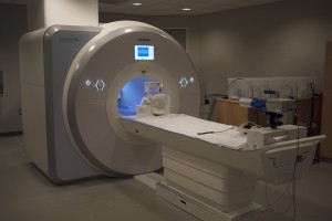
 A lot of experiments in the lab use a tool called Magnetic Resonance Imaging (MRI). This powerful tool allows us to study how the human brain works. An MRI machine (or “scanner”) is essentially a huge and very strong magnet. This magnetic field acts on the atoms in the brain. In particular, it changes how those atoms rotate (or spin). When there’s not a magnetic field, all of the atoms spin in random directions. But in a uniform magnetic field, the atoms spin in the same direction. Then, during an MRI scan, we introduce brief bursts of radio waves (like a radio station) that knock the atoms out of alignment. We measure how many of the waves are resonated back out of the brain as the atoms realign.
To study brain activity and not just anatomy, we use a slight variant called functional MRI (fMRI). The process is basically the same, but we’re looking at a particular molecule, hemoglobin. This molecule transports oxygen through blood vessels in the brain. When an area of the brain is active, it requires a lot of oxygen as fuel, so hemoglobin with oxygen floods the area. When an area is inactive, there is proportionally less oxygenated hemoglobin. Fortunately for us, hemoglobin with and without oxygen has different magnetic properties. So we are able to infer where there has been neural activity.
-Megan deBettencourt
